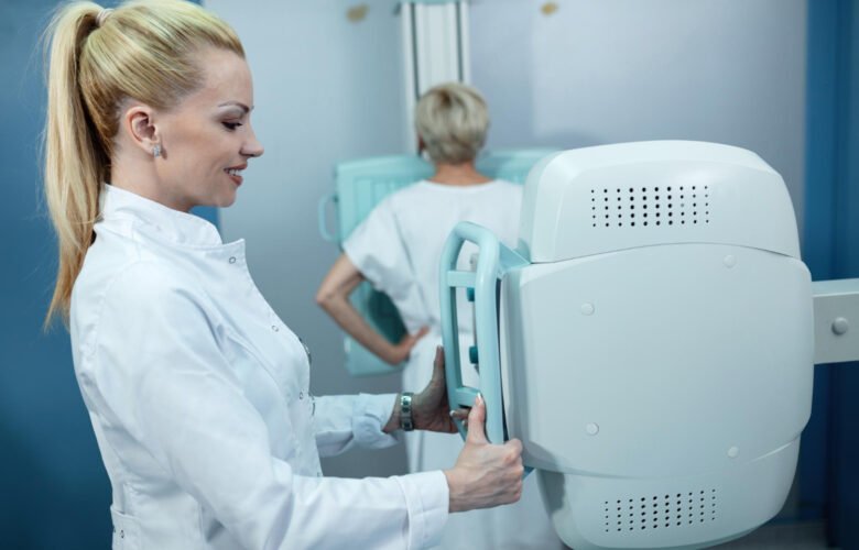
Your mammogram results arrive with reassuring news: no suspicious findings. Below that, another paragraph says you have dense breast tissue. It’s easy to skim past, especially when the main result appears normal.
But that paragraph carries information worth understanding. Since September 10, 2024, mammography facilities covered by federal regulations have been required to notify patients about breast density under updated FDA rules. The standardized language explains that dense tissue can make cancer harder to detect and is associated with increased breast cancer risk.
The notification doesn’t mean something is wrong. It does mean your mammogram result deserves to be understood alongside your individual risk factors.
A Normal Mammogram Doesn’t Tell the Whole Story
Mammograms use X-rays to identify suspicious changes in breast tissue. Fatty tissue generally appears darker, while dense fibroglandular tissue appears white. Some cancers also appear white, making them harder to distinguish from surrounding dense tissue.
The difference becomes particularly relevant in the two highest breast-density categories: heterogeneously dense (C) and extremely dense (D). According to the National Cancer Institute, mammography is less sensitive in dense breasts, meaning some cancers are more likely to go undetected.
A normal mammogram remains valuable, even with dense tissue. But it doesn’t rule out every cancer. If you notice a new lump, nipple discharge, or another concerning breast change, contact your healthcare provider rather than waiting for your next routine screening. Symptoms may require diagnostic evaluation regardless of your most recent mammogram result.
Breast Density Also Changes Your Risk

The difficulty of seeing cancer on a mammogram is only part of the issue. Dense breast tissue is independently associated with a higher likelihood of developing breast cancer compared with predominantly fatty tissue.
That doesn’t mean everyone with dense breasts is at high risk. Age, family history, inherited genetic variants, prior breast biopsies, and other factors influence an individual’s overall risk.
The FDA’s required notification acknowledges both the detection challenge and the increased risk. What it cannot tell you is how those factors combine in your particular situation. That requires a broader discussion with your healthcare provider.
Additional Imaging May Help, but It Isn’t Automatic
Some patients with dense breasts may benefit from supplemental screening beyond mammography. The available options include:
- – Breast MRI. Highly sensitive imaging that is commonly recommended for people at elevated breast cancer risk. It can also produce false-positive findings that require additional testing.
- – Breast ultrasound. An option that may identify some cancers not visible on mammography, although it can lead to unnecessary follow-up imaging or biopsies.
- – Contrast-enhanced mammography. An imaging technique that uses injected contrast material to improve the visibility of certain abnormalities. Availability and suitability vary.
The appropriate choice depends on overall risk, medical history, imaging findings, and patient preferences. Importantly, medical organizations differ on whether supplemental screening should routinely be offered solely because someone has dense breasts.
Additional imaging may find cancers missed by mammography, but it also carries potential harms, including false positives and unnecessary procedures. Insurance coverage varies by plan and state, so patients considering supplemental screening should confirm benefits and ask about appeal options if coverage is denied.
The Follow-Up Conversation Shouldn’t Fall Entirely on You
The density notification is intended to help patients and healthcare professionals make informed decisions. It doesn’t transfer responsibility for appropriate follow-up entirely to the person receiving the letter.
If your results indicate dense tissue, ask your primary care provider, OB-GYN, or breast imaging specialist whether a formal breast cancer risk assessment would be useful. Find out whether your screening schedule is appropriate and whether additional imaging makes sense.
Keep copies of your mammogram letters, imaging reports, and relevant recommendations. If you have concerns about a previous scan or a delayed diagnosis, those records can help establish what was reported, communicated, and recommended.
In some circumstances, a missed cancer may raise questions about whether imaging was properly interpreted or whether appropriate follow-up occurred. A medical malpractice attorney evaluating such a case would need to consider the applicable standard of care, what information was available at the time, and whether any alleged failure caused harm. Dense breast tissue or a delayed diagnosis alone doesn’t establish malpractice.
The paragraph about breast density isn’t a cancer diagnosis or an instruction to schedule every available test. It’s information that can change how you and your healthcare team approach screening. Reading it closely is a good place to start.